CURRENT ISSUE
Watch Online the Live Sessions of ISWWTA 2015 Rishikesh on Youtube.Visit:https://www.youtube.com/user/ayushdarpan/
Previous issues of AYUSH DARPAN in Hindi is now available online visit:http://ayushdarpan.org

Search Engine

Monday, 26 November 2012

Exercise rate related to improvements in Parkinson’s disease


 People with Parkinson's disease benefit from exercise programs on stationary bicycles, with the greatest effect for those who pedal faster, according to a study presented today at the annual meeting of the Radiological Society of North America (RSNA). Functional connectivity magnetic resonance imaging (fcMRI) data showed that faster pedaling led to greater connectivity in brain areas associated with motor ability.
Parkinson's disease is a chronic, progressive disorder of the central nervous system. Early-stage symptoms like shaking and difficulty with walking eventually may progress to cognitive and behavioral problems such as dementia. An estimated 7 to 10 million people worldwide live with Parkinson's disease, according to the Parkinson's Disease Foundation, with most cases occurring after the age of 50.
As the disease progresses and the frequency of side effects increases, the therapeutic window begins to close. Deep brain stimulation is an effective therapy for late-stage Parkinson's disease, but is an invasive and costly procedure.
Exercise is thought to have beneficial effects on Parkinson's disease. Jay L. Alberts, Ph.D., neuroscientist at the Cleveland Clinic Lerner Research Institute in Cleveland, saw this firsthand in 2003 when he rode a tandem bicycle across Iowa with a Parkinson's disease patient to raise awareness of the disease. The patient experienced improvements in her symptoms after the ride.
"The finding was serendipitous," Dr. Alberts recalled. "I was pedaling faster than her, which forced her to pedal faster. She had improvements in her upper extremity function, so we started to look at the possible mechanism behind this improved function."
As part of this inquiry, Dr. Alberts, researcher Chintan Shah, B.S., and their Cleveland Clinic colleagues, recently used fcMRI to study the effect of exercise on 26 Parkinson's disease patients.
"By measuring changes in blood oxygenation levels in the brain, fcMRI allows us to look at the functional connectivity between different brain regions," Shah said.
The patients underwent bicycle exercise sessions three times a week for eight weeks. Some patients exercised at a voluntary level and others underwent forced-rate exercise, pedaling at a speed above their voluntary rate. The researchers used a modified exercise bike to induce forced-rate activity.
"We developed an algorithm to control a motor on the bike and used a controller to sense the patient's rate of exertion and adjust the motor based on their input," Dr. Alberts said.
fcMRI was conducted before and after the eight weeks of exercise therapy and again as follow-up four weeks later. The research team calculated brain activation and connectivity levels from the fcMRI results and correlated the data with average pedaling rate. Results showed increases in task-related connectivity between the primary motor cortex and the posterior region of the brain's thalamus. Faster pedaling rate was the key factor related to these improvements, which were still evident at follow-up.
"The results show that forced-rate bicycle exercise is an effective, low-cost therapy for Parkinson's disease," Shah said.
Dr. Alberts noted that that while faster pedaling led to more significant results, not all Parkinson's patients need to do forced-rate exercise to see improvement.
"We're now looking at this phenomenon in patients with exercise bikes in their home," he said, "and other exercises like swimming and rowing on tandem machines may provide similar benefits."
Source:Radiological Society of North America 

Metabolic protein launches sugar feast that nurtures brain tumors


PKM2 slips into nucleus to promote cancer; potential biomarker and drug approach discovered

 Zhimin Lu, M.D., Ph.D., is an associate professor in MD Anderson's Department of Neuro-Oncology.
 
Researchers at The University of Texas MD Anderson Cancer Center have tracked down a cancer-promoting protein's pathway into the cell nucleus and discovered how, once there, it fires up a glucose metabolism pathway on which brain tumors thrive.
They also found a vital spot along the protein's journey that can be attacked with a type of drug not yet deployed against glioblastoma multiforme, the most common and lethal form of brain cancer. Published online by Nature Cell Biology, the paper further illuminates the importance of pyruvate kinase M2 (PKM2) in cancer development and progression.
"PKM2 is very active during infancy, when you want rapid cell growth, and eventually it turns off. Tumor cells turn PKM2 back on - it's overexpressed in many types of cancer," said Zhimin Lu, M.D., Ph.D., the paper's senior author and an associate professor in MD Anderson's Department of Neuro-Oncology.
Lu and colleagues showed earlier this year that PKM2 in the nucleus also activates a variety of genes involved in cell division. The latest paper shows how it triggers aerobic glycolysis, processing glucose into energy, also known as the Warburg effect, upon which many types of solid tumors rely to survive and grow.
"PKM2 must get to the nucleus to activate genes involved in cell proliferation and the Warburg effect," Lu said. "If we can keep it out of the nucleus, we can block both of those cancer-promoting pathways. PKM2 could be an Achilles' heel for cancer."
By pinpointing the complicated steps necessary for PKM2 to penetrate the nucleus, Lu and colleagues found a potentially druggable target that could keep the protein locked in the cell's cytoplasm.
MEK, ERK emerge as targets
The process begins when the epidermal growth factor connects to its receptor on the cell surface. This leads to:

  • Activation of the MEK protein, which in turn activates ERK.
  • ERK sticking a phosphate group to a specific spot on PKM2.
  • Phosphorylation priming PKM2 for a series of steps that culminate in its binding to the protein importin, which lives up to its name by taking PKM2 through the nuclear membrane.
Once in the nucleus, the team showed that PKM2 activates two genes crucial to aerobic glycolysis and another that splices PKM RNA to make even more PKM2.
An experiment applying several kinase-inhibiting drugs to human glioblastoma cell lines showed that only a MEK/ERK inhibitor prevented EGF-induced smuggling of PKM2 into the nucleus. ERK activation then is mandatory for PKM2 to get into the nucleus.
"MEK/ERK inhibitors have not been tried yet in glioblastoma multiforme," Lu said. Phosporylated PKM2 is a potential biomarker to identify patients who are candidates for MEK/ERK inhibitors once those drugs are developed.
MEK inhibitor blocks tumor growth
The researchers also found that the two glycolysis genes activated by PKM2, called GLUT1 and LDHA, are required for glucose consumption and conversion of pyruvate to lactate, crucial factors in the Warburg Effect. Depleting PKM2 in tumor cell lines reduced glucose consumption and lactate production.
In mice, depleting PKM2 blocked the growth of brain tumors. Re-expressing the wild type protein caused tumors to grow. However, re-expression of a PKM2 mutant protein that lost its ability to get into the nucleus failed to promote tumor formation. Experiments in human glioblastoma cell lines showed the same effect.
Injecting the MEK inhibitor selumetinib into tumors inhibited tumor growth, reduced ERK phosphorylation, PKM2 expression and lactate production in mice. In 48 human tumor samples, the team found that activity of EGFR, ERK1/2 and PKM2 were strongly correlated.
Cause of PKM2 overexpression
Lu and colleagues also published a paper in Molecular Cell that revealed a mechanism for overexpression of PKM2 in glioblastoma. They found that EGF receptor activation turns on NF-?B, which leads to a series of events culminating in PKM2 gene activation.
PKM2 levels were measured in tumor samples from 55 glioblastoma patients treated with standard of care surgery, radiation and chemotherapy. The 20 with low PKM2 expression had a median survival of 34.5 months, compared to 13.6 months for the 35 patients with high levels of PKM2.
Level of PKM2 expression in 27 low-grade astrocytomas was about half of the expression found in higher grade glioblastomas.
"In these two papers, we show how PKM2 is overexpressed in tumors, how it gets into the nucleus, that nuclear entry is essential to tumor development, and identified potential drugs and a biomarker that could usefully treat people," Lu said.
Source:University of Texas M. D. Anderson Cancer Center 

Sunday, 25 November 2012

Union govt allocates Rs.100 cr. for Nano Science and Tech Inst at Mohali, second facility cleared for Karnataka

Union government has set aside Rs.100 crore to establish the country’s first-ever Institute of Nano Science and Technology at Mohali in Punjab. A similar Institute at a cost of Rs.100 crore is also cleared and slated for Bengaluru at Dabaspet Industrial Area on the Peenya-Tumkur Road.In addition, the Centre through the Department of Science and Technology (DST) has pumped in Rs.80 crore to strengthen the nanotechnology infrastructure across the country. To this effect, for setting up the Centres for Nanotechnology or Computational Materials Science (CN/CMS) Karnataka received Rs.25 crore each for the facility at Indian Institute of Science(IISc) and the JNCASR (Jawaharlal Nehru Centre for Advanced Scientific Research). Similar facilities are also underway at Kolkata and Pune.
The CN/CMS would focus on the product or device oriented development but not on basic research only to speed up technology commercialisation. This indicates that India has a come a long way in nanotechnology, stated Prof. CNR Rao, FRS, National Research Professor; Linus Pauling Research Professor; Honorary President, Jawaharlal Nehru Centre for Advanced Scientific Research (JNCASR) and chairman, government of India’s Nano Mission Council and Karnataka Vision Group on Nanotechnology.
The country is ranked fifth globally in nanotechnology R&D with a similar ranking in terms of peer review submissions. Bengaluru has been a pivotal point of action in nanotechnology after Mumbai’s Indian Institute of Technology (IIT) Powai, he added.
For the Institute of Nano Science and Technology, Bengaluru with a Nano Park, the state government has already allocated the land. “Now we need to get the funds from the Centre and in next three to four months and construction will start,” Dr Rao said.
The government of India is looking at the Institute of Nano Science and Technology as specialized centre of excellence to promote the growth of R&D in the areas of nanomaterials, nanometrology, nano-bio pharma, nano-medicine and nano R&D relevant for food and agriculture.
The key objective is to transfer intellectual capital in a bid to maximize the application potential in nanotechnology. Therefore a critical component of the Institute of Nano Science and Technology is human resource development. “We need young scientists with interests in nanotechnology to develop viable products. Start-up companies in India are engaged in designing unique products but what needs to be ensured is feasibility to commercialize the same,” said Dr Rao.
In 2007, government of India spearheaded the Nano Mission only to develop products which have the potential to benefit the country. The technical programmes of the Nano Mission are carried out by the: Nano Science Advisory Group (NSAG) and the Nano Applications and Technology Advisory Group (NATAG).
“The technologically competitive nano science requires ample number of high-quality researchers and therefore the focus of the Nano Mission is to ensure providing valuable education and training in areas of nano-scale science and nano engineering. This is where the Union government’s funding for national institutes and centres will provide the much-needed impetus for growth,” he said.

Source:Pharmabiz

Tea Significantly Reduces Ovarian Cancer Risk

A lifetime of tea drinking can significantly reduce the risk of ovarian cancer in old age, a study has found. 
Health experts carried out a two-year study of 1,000 women with an average age of 59.Half were diagnosed with ovarian cancer while 500 were free of the disease. 
The researchers discovered that those women without cancer were more likely to be tea drinkers from an earlier age than those diagnosed with the illness, the Daily Express reported. 
Flavonoids, powerful compounds with strong disease-fighting properties, were found in black tea in particular, according to the study at the School of Public Health, Curtin University in Perth, Australia. 
"Tea consumption should be encouraged because of the potential benefit in preventing this common and deadly disease," study co-author Dr Andy Lee said. 
Ovarian cancer affects around 7,000 women a year in the UK.
Source-ANI


High Protein Diet May Hasten Wound Healing in Diabetes

French researchers have found that a diet enriched with protein may help to heal wound faster in diabetics. The research offers hope for diabetics who suffer from chronic wounds such as foot ulcers as there is currently no effective way to improve healing of these types of wounds. 
Researchers found that diabetic rats on a high protein diet with arginine and proline—specific molecules found in protein—showed better wound healing over rats fed either standard or high protein food without arginine and proline supplementation.The article is entitled "Arginine plus proline supplementation elicits metabolic adaptation that favors wound healing in diabetic rats." It appears in the online edition of the American Journal of Physiology – Regulatory, Integrative and Comparative Physiology published by the American Physiological Society. 

Methodology 
Researchers divided 18 rats into three groups that were either fed a standard diet, a high-protein diet, or a high protein diet supplemented with arginine and proline (ARG+PRO). On the first day of the experiment, each rat was given an incision, under which a sponge was placed in order to collect wound-healing fluid. To assess skin regrowth and healing, researchers also removed two full-thickness sections of skin from the rats' backs each day from day 1 until day 5, when the experiment ended. 
At the end of the experiment, the rats' blood was analyzed for blood sugar, insulin, and amino acid concentrations. The wounds on their backs were examined for skin regrowth and development of new blood vessels. And, finally, macrophages were collected from the sponges and analyzed for indications of cytokine stimulation and pro-inflammatory activity. 

Results 
Rats on both high protein diets had better nitrogen balance than those on the standard diet. However, the wounds of the rats on the ARG+PRO diet showed more new blood vessel growth on day 5. New blood vessel growth is an essential part of wound healing as the blood vessels supply nutrition and oxygen to growing tissue. 
Furthermore, the macrophages in the ARG+PRO group showed less cytokine stimulation and pro-inflammatory activity than the other groups. This indicates a better environment for promoting wound healing, as inflammation slows the healing process. 
The researchers did not find a difference in skin regrowth between groups, but their findings may be limited because of the small number of rats in the study. Additionally, researchers did not measure markers of collagen deposition in the wound, and the study cannot confirm the beneficial effect of arginine on collagen deposition and wound breaking strength reported in previous research. 

Importance of the Findings 
This study suggests that arginine and proline supplementation could offer new hope for effective treatment in diabetic patients with chronic wounds. This is a promising new area of research where there are no existing effective treatments for these patients. 

Source:American Journal of Physiology 
 

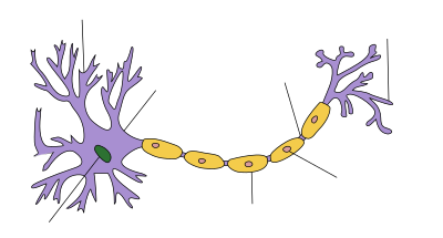
Nerve Cell's Internal Clock Uncovered by Scientists

Scientists have uncovered a nerve cell's internal clock used during embryonic development. This finding offers hope for rejuvenation of damaged brain. The team, led by Dr. Frederic Charron, made the discovery in collaboration with Dr. Alyson Fournier's laboratory at the Montreal Neurological Institute.Researchers in Dr. Charron's laboratory study neurons, which are the nerve cells that make up the central nervous system (brain and spinal cord). They want to better understand how neurons navigate through the developing embryo to arrive at their correct destination. 
"To properly form neural circuits, developing axons (long extensions of neurons that form nerves) follow external signals to reach the right targets," said Dr. Charron, Director of the Molecular Biology of Neural Development research unit at the IRCM. 
"We discovered that nerve cells also have an internal clock, which changes their response to external signals as they develop over time," he stated. 
For this research project, IRCM scientists focused on the Sonic Hedgehog (Shh) protein, which gives cells important information for the embryo to develop properly and plays a critical role in the development of the central nervous system. 
"It is known that axons follow the Shh signal during their development," explained Dr. Patricia Yam, research associate in Dr. Charron's laboratory and first author of the study. 
"However, axons change their behaviour once they reach this protein, and this has been a mystery for the scientific community. We found that a nerve cell's internal clock switches its response to external signals when it reaches the Shh protein, at which time it becomes repelled by the Shh signal rather than following it. 
"Our findings therefore showed that more than one system is involved in directing axon pathfinding during development," said Dr. Yam. 
"Not only do nerve cells respond to external signals, but they also have an internal control system. This discovery is important because it offers new possibilities for developing techniques to regenerate and repair-damaged nerve cells. Along with trying to modify external factors, we can now also consider modifying elements inside a cell in order to change its behaviour," she added. 
Injuries to the central nervous system affect thousands of Canadians every year, and can lead to lifelong disabilities. Most often caused by an accident, stroke or disease, these injuries are very difficult to repair. New tools are therefore required to repair damage to the central nervous system, including techniques that could potentially regenerate nerve cells. 
"The Canadian Institutes of Health Research is delighted to support research aimed at improving the lives of individuals with damage to the brain or spinal cord," said Dr. Anthony Phillips, Scientific Director of CIHR's Institute of Neurosciences, Mental Health and Addiction. 
"Nerve cell repair and regeneration remains an important health challenge, and we believe that Dr. Charron's research findings will contribute to the solution," he added. 
The discovery was published in the prestigious scientific journal Neuron.
Source-ANI
 

India Gets Its Biggest Isotope Labelling Unit

Asia's leading drug discovery research and development organisation, GVK Biosciences, has set up the largest isotope labelling facility in India. 
Isotope labelling is a technique used to track the passage of an isotope, or an atom with a variation, through a reaction.The Hyderabad-based company has also received approvals from the Atomic Energy Regulatory Board (AERB) to carry out 3H and 14C synthesis activities. 
 This Good Laboratory Practice (GLP) compliant facility has state-of-the-art equipment to meet the requirements of companies engaged in the fields of drug discovery and development, agro and veterinary chemicals, cosmetics, perfumery and specialty studies, said a company statement here Monday. 
"Several of our customers requested us to extend our chemistry services to include isotope labelling services. We listened to our customers and set up this state-of-the art facility, the largest such facility in India," said Manni Kantipudi, CEO, GVK BIO. 
GVK BIO's diverse portfolio of nearly 200 customers includes some of the world's largest pharmaceutical, biotechnology, agro, life-sciences companies and leading academic and research institutions.
Source-IANS
   

Facebook Badge

PAGE COUNTER